Category: Eyes & Ears

  • Glaucoma in Dogs: Symptoms, Causes, Treatment, and Prevention

    Home → Dog Health → Eyes & Ears → Glaucoma in Dogs

    Glaucoma in Dogs: Symptoms, Causes, Treatment, and Prevention

    Glaucoma in dogs is a buildup of pressure inside the eye that damages the optic nerve over time, causing pain and, without prompt treatment, permanent vision loss.

    Glaucoma is considered a genuine eye emergency, since the pressure buildup can cause irreversible blindness within hours to days once it becomes severe. This guide covers what causes glaucoma, its symptoms, how it’s diagnosed, how it’s treated, whether it can be prevented, and when to see a veterinarian.

    This guide covers what glaucoma is, what causes it in dogs, its symptoms, how it’s diagnosed, how it’s treated, whether it can be prevented, when to see a vet, and what else people ask.

    What Is Glaucoma in Dogs?

    Glaucoma is an increase in pressure inside the eye that occurs when fluid produced inside the eye doesn’t drain properly, causing pressure to build and gradually damage the optic nerve, the structure responsible for carrying visual information to the brain. Veterinarians classify glaucoma as primary, meaning it develops independently, often with a genetic basis, or secondary, meaning it results from another eye condition that interferes with normal fluid drainage. Both forms are serious and can lead to permanent blindness in the affected eye without prompt treatment.

    What Causes Glaucoma in Dogs?

    Primary glaucoma has a genetic basis in many affected breeds, including the Cocker Spaniel, Basset Hound, and Chow Chow, and typically results from an inherited abnormality in the eye’s fluid drainage structures. Secondary glaucoma develops as a complication of another eye problem, including chronic uveitis, an advanced cataract, a dislocated lens, or a tumor inside the eye, all of which can physically block normal fluid drainage. Because the two forms have such different underlying causes, identifying which type a dog has shapes both treatment and the long-term outlook, including the risk to the other eye.

    What Are the Symptoms of Glaucoma in Dogs?

    Redness, a cloudy or bluish appearance to the cornea, and visible signs of pain, including squinting, pawing at the eye, or unusual lethargy, are common early symptoms of glaucoma. A dilated pupil that doesn’t respond normally to light is another important sign, and as pressure continues building, the affected eye can begin to look visibly larger than normal, a change called buphthalmos that develops in longstanding, chronic cases. Vision loss in the affected eye can happen gradually or, in an acute glaucoma crisis, within a matter of hours.

    How Is Glaucoma in Dogs Diagnosed?

    A veterinarian diagnoses glaucoma using tonometry, a specialized instrument that measures the pressure inside the eye directly, which is the single most important test for confirming the condition. A complete eye exam helps determine whether the glaucoma is primary or secondary by looking for signs of another underlying eye problem that might be driving the pressure increase. Because glaucoma can progress so quickly, same-day diagnosis and treatment matter considerably once the condition is suspected, and referral to a veterinary ophthalmologist is common given the specialized equipment and expertise involved.

    How Is Glaucoma in Dogs Treated?

    Glaucoma is treated as a genuine emergency, with medication aimed at rapidly lowering the eye’s internal pressure to prevent further damage to the optic nerve. Depending on the cause and how the eye responds to initial medical treatment, a veterinary ophthalmologist may recommend surgery to improve fluid drainage or reduce fluid production inside the eye. In cases where an eye has already lost vision and remains chronically painful despite treatment, removing the eye, a procedure called enucleation, is sometimes recommended to permanently relieve pain, and because primary glaucoma often eventually affects both eyes, the unaffected eye is typically monitored closely and sometimes treated preventively.

    Can Glaucoma in Dogs Be Prevented?

    Primary glaucoma generally can’t be prevented, since it stems from an inherited structural predisposition, though regular eye pressure screening in predisposed breeds can catch the condition early, before it causes significant, irreversible damage. Secondary glaucoma is sometimes preventable by promptly and thoroughly treating the underlying eye condition that could otherwise progress to cause it, including chronic uveitis or an unmanaged cataract. For a dog that has already developed primary glaucoma in one eye, preventive monitoring and treatment of the other eye can meaningfully delay or reduce the severity of glaucoma developing there as well.

    When Should You See a Vet for Glaucoma in Dogs?

    Seek emergency veterinary care immediately if a dog’s eye becomes suddenly red, cloudy, or painful, or if the pupil looks unusually large or unresponsive to light, since these are classic signs of a glaucoma crisis requiring same-day treatment. Any dog from a breed predisposed to primary glaucoma should have routine eye pressure checks as part of regular veterinary care, since early detection meaningfully improves the chances of preserving vision. A dog already diagnosed with glaucoma in one eye needs ongoing, close monitoring of the other eye given the real risk of it developing the condition as well.

    What Else Do People Ask About Glaucoma in Dogs?

    The questions below cover whether glaucoma is curable, how quickly it can cause blindness, whether glaucoma is painful, and which dogs are most at risk.

    Is glaucoma in dogs curable?

    No, glaucoma is not curable, and any vision already lost to optic nerve damage cannot be restored. Treatment focuses on controlling eye pressure to prevent further damage and relieve pain, and the earlier treatment starts, the better the chances of preserving remaining vision.

    How quickly can glaucoma cause blindness in dogs?

    An acute glaucoma crisis can cause permanent blindness in the affected eye within as little as a day if pressure isn’t brought down quickly, which is exactly why glaucoma is treated as a true eye emergency. Chronic, slowly progressing glaucoma causes vision loss more gradually but still carries a serious long-term risk to sight.

    Is glaucoma painful for dogs?

    Yes, glaucoma is genuinely painful, since the elevated pressure inside the eye directly affects sensitive internal structures. Dogs often show signs of this pain, including squinting, pawing at the eye, lethargy, or reduced appetite, even when the discomfort isn’t immediately obvious to an owner.

    Which dogs are most at risk for glaucoma?

    Breeds including the Cocker Spaniel, Basset Hound, and Chow Chow show a well-documented genetic predisposition to primary glaucoma, while any dog with a chronic eye condition, including uveitis or advanced cataracts, faces an elevated risk of secondary glaucoma. A veterinarian can advise on appropriate screening based on an individual dog’s breed and health history.

    Related Guides

    More guides on The Dogs Wiki cover general dog health and senior dog care.

    Guide
    Dog Health
  • Ear Infections in Dogs: Symptoms, Causes, Treatment, and Prevention

    Home → Dog Health → Eyes & Ears → Ear Infections in Dogs

    Ear Infections in Dogs: Symptoms, Causes, Treatment, and Prevention

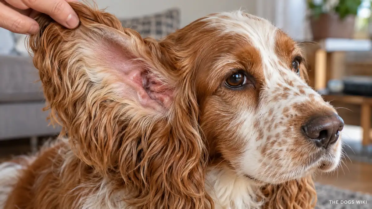
    Ear infections in dogs, medically called otitis externa, are inflammation and infection of the ear canal, most often caused by an overgrowth of bacteria or yeast that’s normally present in small amounts.

    Dogs with floppy ears or a history of allergies face a considerably higher risk of recurring ear infections, since both trap moisture or disrupt the ear’s normal defenses against bacterial and yeast overgrowth. This guide covers what causes ear infections, their symptoms, how they’re diagnosed, how they’re treated, whether they can be prevented, and when to see a veterinarian.

    This guide covers what an ear infection is, what causes it in dogs, its symptoms, how it’s diagnosed, how it’s treated, whether it can be prevented, when to see a vet, and what else people ask.

    What Is an Ear Infection in Dogs?

    An ear infection in dogs is inflammation of the ear canal caused by an overgrowth of bacteria, yeast, or sometimes both, organisms that exist in small, normal numbers in a healthy ear but multiply out of control when the ear’s environment changes. The condition is one of the most common reasons dogs are brought to the veterinarian, and it ranges from a mild, easily treated case to a chronic, recurring problem requiring ongoing management. Left untreated, a bacterial or yeast ear infection can progress to involve the middle or inner ear, which raises the risk of more serious complications, including balance problems.

    What Causes Ear Infections in Dogs?

    Moisture trapped in the ear canal, whether from swimming, bathing, or humid weather, creates an environment where bacteria and yeast multiply more easily than they would in a dry ear. Allergies, whether environmental or food-related, are one of the most significant underlying drivers of recurring ear infections, since allergic inflammation changes the ear canal’s environment in a way that favors overgrowth. Breeds with floppy ears face an elevated risk since their ear shape restricts airflow, and other contributing factors include ear mites, foreign objects lodged in the canal, and excessive hair growth inside the ear canal in certain breeds.

    What Are the Symptoms of Ear Infections in Dogs?

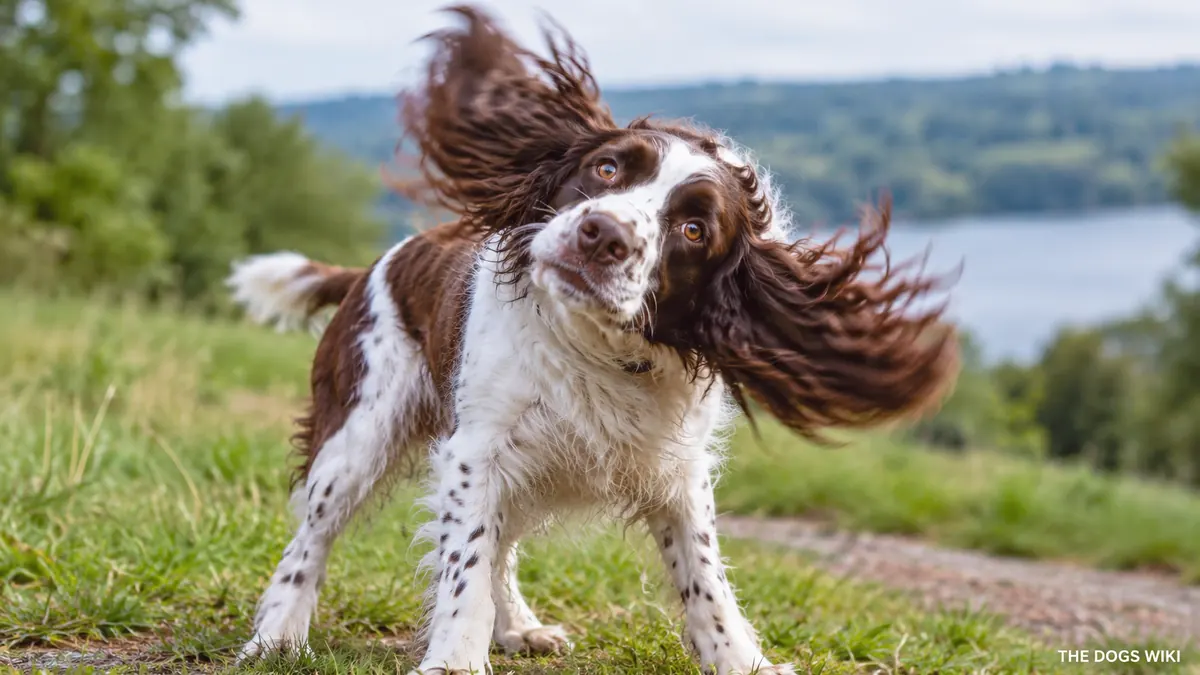
    Head shaking and scratching at the affected ear are typically the earliest and most noticeable symptoms, reflecting the itching and discomfort an infection causes. A noticeable odor and visible discharge, which can range from dark and waxy to yellow or pus-like, are also common, along with redness and swelling inside the visible part of the ear canal. In more painful or advanced infections, a dog may resist having the ear touched, tilt its head to one side, or seem generally less interested in normal activity.

    How Are Ear Infections in Dogs Diagnosed?

    A veterinarian diagnoses an ear infection through a visual exam using an otoscope, which allows a direct look into the ear canal to assess redness, discharge, and whether the eardrum is intact. Ear cytology, in which a sample of the discharge is examined under a microscope, identifies whether bacteria, yeast, or both are present and helps guide the choice of treatment. For dogs with recurring infections, a veterinarian often recommends additional testing to identify an underlying cause, such as allergy testing, since treating only the infection without addressing the root cause leads to repeat problems.

    How Are Ear Infections in Dogs Treated?

    Treatment typically starts with a thorough ear cleaning to remove discharge and debris, which also helps any subsequent medication reach the affected tissue more effectively. A veterinarian then prescribes a topical medication matched to the specific cause identified through cytology, commonly an antibiotic for bacterial infections, an antifungal for yeast infections, or a combination product along with an anti-inflammatory to reduce swelling and discomfort. For dogs with allergy-driven recurring infections, managing the underlying allergy, whether through dietary changes, environmental control, or allergy medication, is an essential part of breaking the cycle of repeat infections.

    Can Ear Infections in Dogs Be Prevented?

    Keeping ears clean and dry, particularly after swimming or bathing, reduces the moisture that bacteria and yeast need to multiply, and routine ear checks catch early signs of infection before they become more serious. Managing an underlying allergy with a veterinarian’s guidance considerably reduces the frequency of infections in allergy-prone dogs. Breeds with floppy ears or excessive ear hair benefit from more frequent preventive ear care, including regular cleaning as part of a routine grooming schedule.

    When Should You See a Vet for Ear Infections in Dogs?

    See a veterinarian if a dog is shaking its head, scratching at an ear, or showing visible redness, odor, or discharge, since prompt treatment prevents the infection from worsening or spreading further into the ear. A dog tilting its head, losing balance, or showing signs of significant pain needs a more urgent evaluation, since these can indicate the infection has progressed to affect the middle or inner ear. Dogs with a history of recurring ear infections should see a veterinarian for a broader workup rather than treating each episode in isolation, since an underlying cause is likely driving the pattern.

    What Else Do People Ask About Ear Infections in Dogs?

    The questions below cover whether ear infections are contagious, how often floppy-eared dogs need ear cleaning, whether ear infections resolve on their own, and whether allergies are really connected to ear infections.

    Are ear infections in dogs contagious?

    No, most ear infections in dogs are not contagious between dogs, since they typically result from an overgrowth of organisms already present in that individual dog’s ear rather than an organism transmitted from another animal. Ear mites are an exception and can spread between dogs in close contact, which is one reason a veterinary diagnosis matters for identifying the specific cause.

    How often should floppy-eared dogs have their ears cleaned?

    Floppy-eared breeds generally benefit from ear cleaning roughly weekly to every two weeks, though a veterinarian can recommend a specific schedule based on an individual dog’s ear shape and infection history. Over-cleaning can sometimes irritate the ear canal, so following a veterinarian’s specific guidance matters more than cleaning as often as possible.

    Can an ear infection in dogs resolve on its own?

    No, ear infections in dogs generally don’t resolve without treatment and tend to worsen if left unaddressed, since the underlying bacterial or yeast overgrowth typically continues unchecked. Prompt veterinary treatment prevents the discomfort and potential complications of a worsening infection.

    Are allergies really connected to ear infections in dogs?

    Yes, allergies are one of the most significant underlying causes of recurring ear infections in dogs, since allergic inflammation changes the ear canal environment in ways that favor bacterial and yeast overgrowth. A dog with repeated ear infections and no other obvious cause is often a good candidate for allergy evaluation.

    Related Guides

    More guides on The Dogs Wiki cover general dog health and grooming.

    Guide
    Dog Health
    Grooming
  • Conjunctivitis in Dogs: Symptoms, Causes, Treatment, and Prevention

    Home → Dog Health → Eyes & Ears → Conjunctivitis in Dogs

    Conjunctivitis in Dogs: Symptoms, Causes, Treatment, and Prevention

    Conjunctivitis in dogs is inflammation of the conjunctiva, the pink membrane lining the inside of the eyelid and covering the white part of the eye, and it can stem from allergies, infection, irritation, or an underlying eye problem.

    Because conjunctivitis can look similar regardless of its underlying cause, figuring out exactly what’s triggering a specific dog’s inflamed eye matters for choosing the right treatment rather than guessing. This guide covers what causes conjunctivitis, its symptoms, how it’s diagnosed, how it’s treated, whether it can be prevented, and when to see a veterinarian.

    This guide covers what conjunctivitis is, what causes it in dogs, its symptoms, how it’s diagnosed, how it’s treated, whether it can be prevented, when to see a vet, and what else people ask.

    What Is Conjunctivitis in Dogs?

    Conjunctivitis is inflammation of the conjunctiva, the thin, pink membrane that lines the inner surface of the eyelids and covers the white part of the eye. This inflammation causes the characteristic redness and swelling associated with the condition, and it can affect one or both eyes depending on the underlying cause. Conjunctivitis itself is often described as a symptom as much as a standalone diagnosis, since a range of different underlying problems can all produce this same basic inflammatory response in the eye.

    What Causes Conjunctivitis in Dogs?

    Allergies, whether environmental or seasonal, are a common cause of conjunctivitis, producing inflammation as part of a broader allergic response that can also affect the skin and ears. Bacterial and viral infections can directly infect the conjunctiva and cause inflammation, often producing a thicker, colored discharge than allergy-driven cases typically show. Environmental irritants, including dust, smoke, and foreign material like grass seeds, can trigger conjunctivitis mechanically, and underlying eye conditions, including dry eye or eyelid abnormalities that cause chronic irritation, can also produce ongoing or recurring conjunctivitis.

    What Are the Symptoms of Conjunctivitis in Dogs?

    Redness and swelling of the tissue around the eye are the hallmark symptoms of conjunctivitis, often accompanied by some degree of discharge that can range from clear and watery in allergic or irritant cases to thick and yellow-green in bacterial infections. Many affected dogs squint, paw at the affected eye, or show general discomfort, and the eye may appear partially closed if the swelling and irritation are significant. Excessive blinking and sensitivity to light can also accompany more severe cases.

    How Is Conjunctivitis in Dogs Diagnosed?

    A veterinarian diagnosing conjunctivitis performs a thorough eye exam to assess the pattern and severity of inflammation and look for any foreign material or structural abnormality that might be contributing. A fluorescein stain test, which highlights any damage to the surface of the eye, helps rule out a corneal ulcer, a different and potentially more serious condition that can accompany or mimic conjunctivitis. Depending on the suspected cause, a veterinarian may also recommend a tear production test or a sample of the discharge to identify a specific infectious organism.

    How Is Conjunctivitis in Dogs Treated?

    Treatment for conjunctivitis depends entirely on its underlying cause: bacterial infections are treated with antibiotic eye drops or ointment, while allergic conjunctivitis responds to anti-inflammatory or antihistamine treatment alongside managing the broader allergy if one is identified. A foreign object or irritant lodged against the eye is flushed out, which often resolves the irritation once the offending material is removed. For conjunctivitis linked to an underlying structural eye problem, such as an eyelid abnormality, addressing that root cause is necessary to prevent the conjunctivitis from recurring.

    Can Conjunctivitis in Dogs Be Prevented?

    Managing a known allergy with a veterinarian’s guidance reduces the frequency of allergy-related conjunctivitis flare-ups considerably. Keeping a dog’s environment reasonably free of irritants, including excessive dust or smoke exposure, and promptly removing visible debris from around the eyes after outdoor activity both help reduce irritant-driven cases. Addressing any underlying structural eye issue identified by a veterinarian also prevents the chronic, recurring conjunctivitis that condition can otherwise cause.

    When Should You See a Vet for Conjunctivitis in Dogs?

    See a veterinarian if a dog’s eye becomes red, swollen, or starts producing discharge, since an accurate diagnosis determines the right treatment and rules out more serious conditions that can look similar. Seek prompt veterinary care if a dog is squinting significantly, pawing persistently at the eye, or showing signs of real pain, since these can indicate a more serious problem, including a corneal ulcer, rather than straightforward conjunctivitis. Any eye symptom that doesn’t improve within a day or two of starting home observation, or that worsens, warrants a veterinary evaluation rather than continued waiting.

    What Else Do People Ask About Conjunctivitis in Dogs?

    The questions below cover whether conjunctivitis is contagious, whether it can be treated at home, how long it typically lasts, and whether conjunctivitis can affect a dog’s vision.

    Is conjunctivitis in dogs contagious?

    Conjunctivitis caused by certain bacterial or viral infections can potentially spread to other dogs in close contact, while allergy or irritant-driven conjunctivitis is not contagious at all. A veterinarian can advise on whether isolation from other pets is warranted based on the specific cause identified.

    Can conjunctivitis in dogs be treated at home?

    No, conjunctivitis generally needs a proper veterinary diagnosis before treatment, since using the wrong type of eye medication, or treating a corneal ulcer as simple conjunctivitis, can make the underlying problem worse. A veterinarian can prescribe the right medication once the specific cause is identified.

    How long does conjunctivitis typically last in dogs?

    Most cases of conjunctivitis improve within a week or two with appropriate treatment targeted at the underlying cause, though allergy-related cases can recur whenever the dog is exposed to the trigger again. Conjunctivitis linked to an untreated structural eye issue can persist or keep recurring until that root cause is addressed.

    Can conjunctivitis affect a dog’s vision?

    Uncomplicated conjunctivitis generally doesn’t cause lasting vision problems once properly treated, since the inflammation is limited to the membrane surrounding the eye rather than the structures responsible for sight. Vision can be affected if conjunctivitis is actually masking or accompanying a more serious condition, which is part of why a proper veterinary exam matters rather than assuming it’s a minor issue.

    Related Guides

    More guides on The Dogs Wiki cover general dog health.

    Guide
    Dog Health
  • Cherry Eye in Dogs: Symptoms, Causes, Treatment, and Prevention

    Home → Dog Health → Eyes & Ears → Cherry Eye in Dogs

    Cherry Eye in Dogs: Symptoms, Causes, Treatment, and Prevention

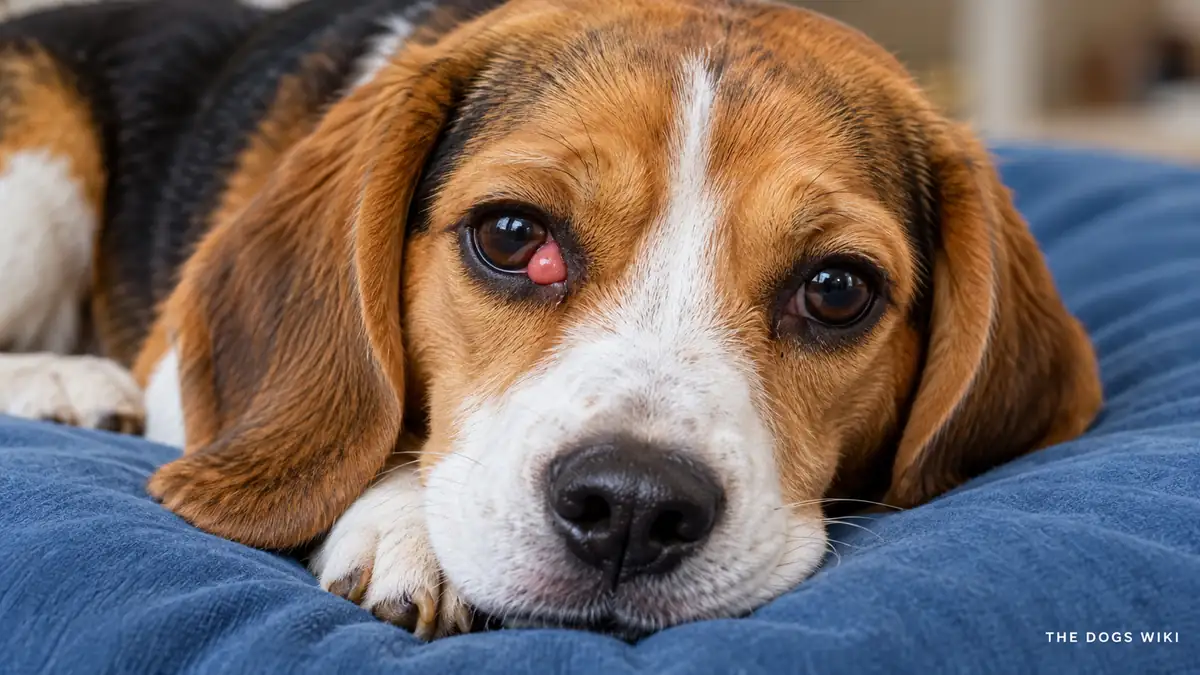
    Cherry eye in dogs is the prolapse of a gland located in the third eyelid, appearing as a reddish, round swelling in the inner corner of the eye, and it’s considerably more common in certain breeds.

    Despite its alarming appearance, cherry eye isn’t typically painful in its early stages, though the condition needs prompt treatment to protect the gland’s important role in tear production. This guide covers what causes cherry eye, its symptoms, how it’s diagnosed, how it’s treated, whether it can be prevented, and when to see a veterinarian.

    This guide covers what cherry eye is, what causes it in dogs, its symptoms, how it’s diagnosed, how it’s treated, whether it can be prevented, when to see a vet, and what else people ask.

    What Is Cherry Eye in Dogs?

    Cherry eye is the common name for prolapse of the gland of the third eyelid, a structure located at the inner corner of a dog’s eye that normally sits unseen and contributes a meaningful portion of the eye’s tear film. When the connective tissue holding this gland in place weakens, the gland flips up and becomes visible as a round, reddish mass, which is where the condition gets its descriptive nickname. The condition can affect one or both eyes and most often appears in young dogs.

    What Causes Cherry Eye in Dogs?

    Cherry eye results from a weakness in the connective tissue that normally anchors the third eyelid gland in place, allowing the gland to flip outward and become visible. The exact reason this connective tissue weakens isn’t fully understood, but genetics plays a clear role, since certain breeds, including the Bulldog, Cocker Spaniel, Beagle, Shih Tzu, Lhasa Apso, and Bloodhound, show a well-documented predisposition to the condition. Cherry eye most commonly appears in dogs under two years old, suggesting a developmental component to the underlying weakness.

    What Are the Symptoms of Cherry Eye in Dogs?

    The defining symptom of cherry eye is a visible, round, reddish-pink mass at the inner corner of the affected eye, often appearing somewhat suddenly. The gland itself isn’t usually painful early on, though a dog may paw at the eye or show mild irritation as the exposed tissue becomes dry or irritated from exposure. Left untreated for an extended period, the exposed gland can become increasingly inflamed, swollen, and prone to drying out, which raises the risk of longer-term complications affecting tear production.

    How Is Cherry Eye in Dogs Diagnosed?

    Cherry eye is typically diagnosed through a straightforward visual exam, since the prolapsed gland’s appearance is distinctive and recognizable to a veterinarian. A broader eye exam helps rule out other conditions that could coexist with or mimic cherry eye, and a veterinarian also checks the overall health of the eye and surrounding tissue to plan the most appropriate treatment approach. No advanced imaging or lab testing is typically needed to confirm a cherry eye diagnosis.

    How Is Cherry Eye in Dogs Treated?

    Surgical repositioning of the gland, rather than removal, is the standard and strongly recommended treatment for cherry eye, since the gland plays an important role in producing tears and removing it entirely raises the long-term risk of dry eye. Several surgical techniques exist for securing the gland back into its normal position, and a veterinary ophthalmologist or experienced veterinarian can determine which approach fits a specific case best. Prompt surgical treatment generally produces good outcomes and preserves normal tear production going forward.

    Can Cherry Eye in Dogs Be Prevented?

    Cherry eye generally can’t be prevented in a dog with the underlying genetic predisposition for weak connective tissue in this area, since it’s a structural issue rather than something caused by an external factor an owner can control. Responsible breeders can help reduce the condition’s prevalence over generations by avoiding breeding dogs that have experienced cherry eye, particularly within predisposed breeds. For an individual affected dog, prompt treatment is the most effective way to prevent the complications that can develop if the condition goes unaddressed.

    When Should You See a Vet for Cherry Eye in Dogs?

    See a veterinarian promptly if you notice a reddish, round mass at the inner corner of a dog’s eye, since early treatment generally leads to better outcomes and lower risk of complications. It’s worth scheduling the visit soon rather than waiting, even though the condition isn’t usually an emergency in its early stages, since prolonged exposure of the gland raises the risk of lasting damage to tear production. A dog that’s already had cherry eye in one eye should also be watched for the same issue developing in the other eye, since the condition can affect both over time.

    What Else Do People Ask About Cherry Eye in Dogs?

    The questions below cover whether cherry eye is painful, whether it can go away without surgery, which dogs are most at risk, and whether cherry eye can come back after surgery.

    Is cherry eye painful for dogs?

    Cherry eye isn’t typically painful in its early stages, though a dog may show mild irritation or paw at the eye as the exposed gland dries out or becomes inflamed over time. Leaving the condition untreated for an extended period increases the likelihood of discomfort developing as complications set in.

    Can cherry eye go away without surgery?

    No, cherry eye generally doesn’t resolve on its own and requires surgical correction to reposition the gland properly. Some owners try massage or other non-surgical approaches, but these aren’t considered reliable solutions and surgery remains the standard, recommended treatment.

    Which dogs are most at risk for cherry eye?

    Breeds including the Bulldog, Cocker Spaniel, Beagle, Shih Tzu, Lhasa Apso, and Bloodhound show a well-documented elevated risk for cherry eye, and the condition most often appears in dogs under two years old. Any breed can potentially develop cherry eye, but these breeds account for a disproportionate share of diagnosed cases.

    Can cherry eye come back after surgery?

    Yes, cherry eye can recur after surgery in some dogs, though modern surgical techniques that reposition rather than remove the gland have generally improved success rates considerably. A veterinarian can discuss the specific recurrence risk associated with the surgical technique used for an individual dog.

    Related Guides

    More guides on The Dogs Wiki cover general dog health.

    Guide
    Dog Health
  • Cataracts in Dogs: Symptoms, Causes, Treatment, and Prevention

    Home → Dog Health → Eyes & Ears → Cataracts in Dogs

    Cataracts in Dogs: Symptoms, Causes, Treatment, and Prevention

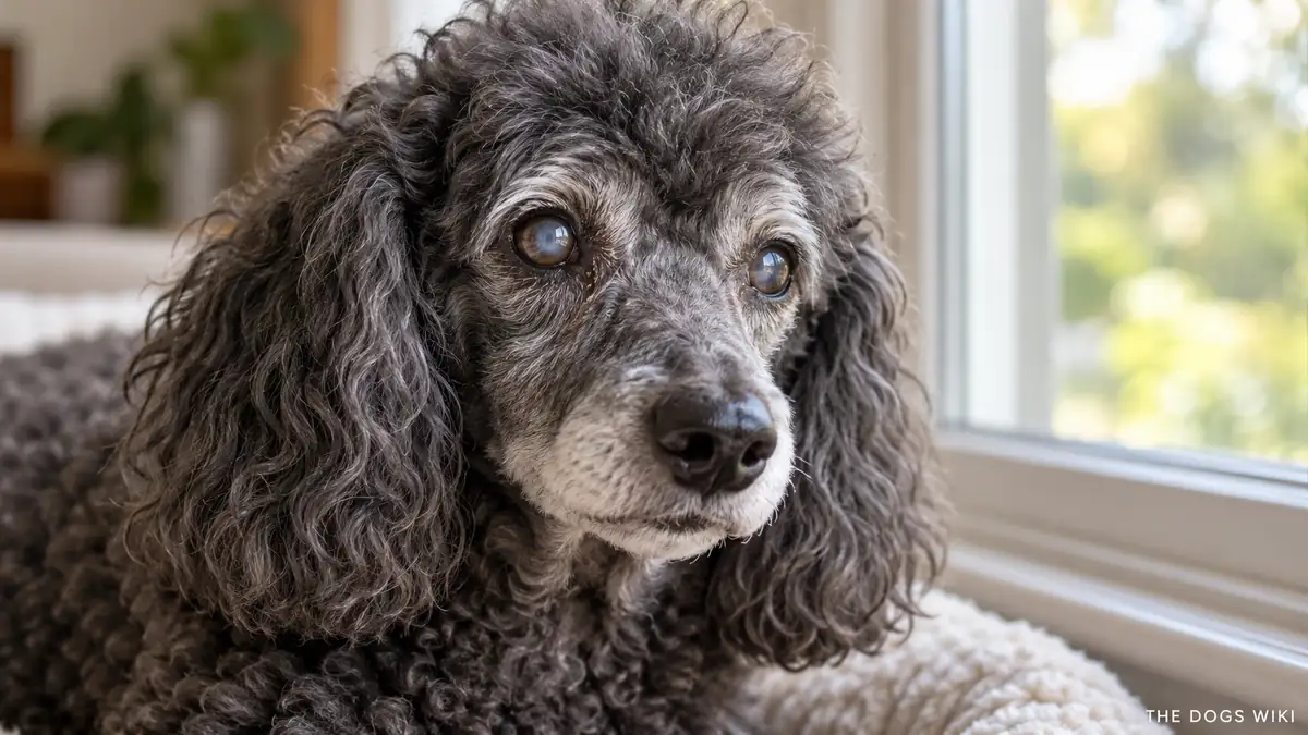
    Cataracts in dogs are a clouding of the eye’s lens that interferes with light passing through to the retina, gradually or sometimes rapidly reducing vision and, in advanced cases, causing complete blindness.

    Diabetes is one of the strongest known risk factors for cataracts in dogs, with many diabetic dogs developing significant lens clouding within months of their diabetes diagnosis, which is one of several reasons cataracts deserve a proper veterinary workup rather than being treated as a simple age-related nuisance. This guide covers what causes cataracts, their symptoms, how they’re diagnosed, how they’re treated, whether they can be prevented, and when to see a veterinarian.

    This guide covers what cataracts are, what causes them in dogs, their symptoms, how they’re diagnosed, how they’re treated, whether they can be prevented, when to see a vet, and what else people ask.

    What Are Cataracts in Dogs?

    Cataracts are an opacity, or clouding, that develops in the lens of the eye, the structure responsible for focusing light onto the retina to produce clear vision. As a cataract progresses, it blocks more light from passing through, gradually reducing vision and eventually causing blindness in the affected eye if the entire lens becomes opaque. Cataracts can affect one or both eyes, develop at any age depending on the underlying cause, and range from a small, vision-sparing spot to complete, vision-obscuring lens opacity.

    What Causes Cataracts in Dogs?

    Inherited or genetic cataracts are common in dogs, with many breeds showing a documented predisposition to developing them, sometimes even at a young age in a form called juvenile cataracts. Diabetes is one of the most significant and well-documented causes of cataracts in dogs, since the high blood sugar associated with the disease alters the lens’s internal chemistry in a way that frequently causes rapid cataract formation. Age-related cataracts develop gradually in many older dogs as a natural part of the aging process, and trauma to the eye can also directly damage the lens and trigger cataract formation.

    What Are the Symptoms of Cataracts in Dogs?

    A visibly cloudy, white, or bluish-gray appearance to the pupil is the most recognizable sign of cataracts, though the exact appearance varies depending on how much of the lens is affected. As vision declines, owners often notice behavioral changes, including increased hesitancy on stairs, bumping into furniture, particularly in dim lighting, or a general reluctance to navigate unfamiliar spaces. Early, small cataracts may not produce any noticeable vision changes at all and are sometimes found incidentally during a routine eye exam.

    How Are Cataracts in Dogs Diagnosed?

    A veterinarian diagnoses cataracts through a direct eye exam, often using specialized equipment to get a detailed view of the lens and assess how much of it is affected. Because cataracts and the normal, age-related lens haziness called nuclear sclerosis can look somewhat similar to an untrained eye, a veterinary exam is important for an accurate diagnosis rather than assuming cloudiness is simply a sign of normal aging. Given the strong link between diabetes and cataracts, a veterinarian typically recommends bloodwork to check blood sugar levels in any dog newly diagnosed with cataracts, particularly if the cataracts appeared and progressed quickly.

    How Are Cataracts in Dogs Treated?

    Surgical removal of the cataract, a procedure called phacoemulsification performed by a veterinary ophthalmologist, is currently the only treatment that can restore vision lost to cataracts, and it involves removing the clouded lens and typically replacing it with an artificial one. Cataracts that aren’t surgically treated don’t resolve on their own and can lead to secondary complications over time, including inflammation inside the eye and an elevated risk of glaucoma. For dogs that aren’t good candidates for surgery, due to other health conditions or owner preference, a veterinarian can discuss managing the eye to reduce complications even though vision won’t be restored.

    Can Cataracts in Dogs Be Prevented?

    Cataracts caused by inherited genetic factors generally can’t be prevented, since the predisposition is built into the dog’s genetics from birth. Diabetes-related cataracts can sometimes be slowed or reduced in severity through excellent blood sugar control, which is one of several reasons consistent management of diabetes matters beyond the disease itself. Protecting a dog’s eyes from trauma and scheduling regular veterinary eye exams, particularly for predisposed breeds, help catch cataracts early even when they can’t be prevented outright.

    When Should You See a Vet for Cataracts in Dogs?

    See a veterinarian if you notice any cloudiness, color change, or unusual appearance in a dog’s eye, or if a dog seems to be struggling with vision, bumping into objects, or hesitant in situations it previously navigated easily. A dog with diagnosed diabetes should have its eyes checked regularly, given how commonly and quickly diabetic cataracts can develop. A sudden onset of significant eye redness, pain, or vision loss alongside existing cataracts warrants urgent veterinary attention, since this combination can indicate a serious complication like glaucoma developing.

    What Else Do People Ask About Cataracts in Dogs?

    The questions below cover whether cataracts are reversible without surgery, how quickly cataracts progress, whether cataract surgery is worth it for an older dog, and whether cataracts are painful.

    Are cataracts in dogs reversible without surgery?

    No, cataracts don’t reverse or resolve without surgery, since the lens clouding is a structural change that doesn’t spontaneously clear on its own. Surgical removal remains the only treatment currently available to restore vision lost to cataracts.

    How quickly do cataracts progress in dogs?

    Progression speed varies considerably depending on the underlying cause, with diabetes-related cataracts sometimes developing to full opacity within days to weeks, while age-related or some inherited cataracts progress much more gradually over months to years. A veterinarian can offer a more specific sense of expected progression based on the type and cause identified in an individual dog.

    Is cataract surgery worth it for an older dog?

    Cataract surgery can be worth it for an older dog in good overall health, since age alone isn’t typically a barrier to successful surgery and many older dogs benefit meaningfully from restored vision. A veterinary ophthalmologist evaluates a dog’s overall health and the eye’s suitability for surgery to help an owner make this decision.

    Are cataracts painful for dogs?

    Cataracts themselves generally aren’t painful, though associated complications, including inflammation or secondary glaucoma, can cause real discomfort if they develop. A veterinarian can assess whether any pain a dog with cataracts is showing stems from the cataract itself or a related complication.

    Related Guides

    More guides on The Dogs Wiki cover general dog health and senior dog care.

    Guide
    Dog Health