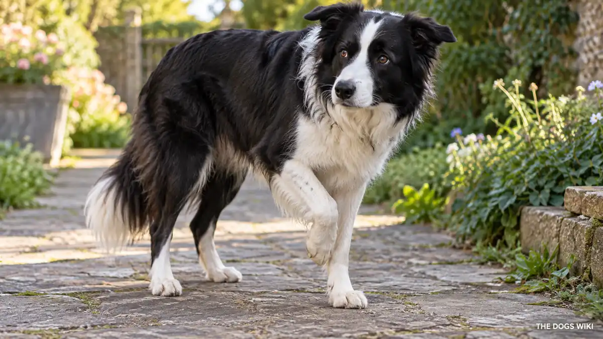
Category: Orthopedic & Joint

  • Torn ACL (Cruciate Ligament) in Dogs: Symptoms, Causes, Treatment, and Prevention

    Home → Dog Health → Orthopedic & Joint → Torn ACL (Cruciate Ligament) in Dogs

    Torn ACL (Cruciate Ligament) in Dogs: Symptoms, Causes, Treatment, and Prevention

    A torn ACL in dogs, more precisely called a cranial cruciate ligament rupture, is damage to the ligament that stabilizes the knee joint, and it’s one of the most common orthopedic injuries veterinarians treat.

    Unlike the sudden, trauma-driven ACL tears common in human athletes, most cruciate ligament injuries in dogs result from gradual degeneration of the ligament over time, which is why the injury can sometimes seem to happen with minimal or no obvious trauma. This guide covers what causes a torn cruciate ligament, its symptoms, how it’s diagnosed, how it’s treated, whether it can be prevented, and when to see a veterinarian.

    This guide covers what a torn ACL is, what causes it in dogs, its symptoms, how it’s diagnosed, how it’s treated, whether it can be prevented, when to see a vet, and what else people ask.

    What Is a Torn ACL in Dogs?

    A torn ACL, medically called a cranial cruciate ligament rupture, is damage to the ligament inside the knee joint responsible for preventing the shin bone from sliding forward relative to the thigh bone. This ligament is the canine equivalent of the human anterior cruciate ligament, though the two species’ knee anatomy and the typical mechanism of injury differ somewhat. Once this ligament is torn or significantly weakened, the knee joint becomes unstable, which causes pain, lameness, and, over time, the development of arthritis within the joint.

    What Causes a Torn ACL in Dogs?

    Gradual degeneration of the ligament over time is the most common underlying cause in dogs, a process influenced by genetics, chronic low-grade inflammation within the joint, and the dog’s individual knee conformation, which eventually weakens the ligament enough that it tears, sometimes during normal activity. Excess body weight significantly increases the stress placed on the ligament and accelerates this degenerative process. Sudden traumatic injury, including a hard landing or a quick twisting motion, can also cause a cruciate ligament tear, particularly in an active, athletic dog, though this is a less common cause overall than gradual degeneration.

    What Are the Symptoms of a Torn ACL in Dogs?

    Sudden or gradually worsening hind-limb lameness is the hallmark symptom of a torn cruciate ligament, ranging from a subtle limp to a complete refusal to bear weight on the affected leg. Swelling around the knee joint is common, and many affected dogs sit with the affected leg held out to the side rather than tucked normally underneath them. Because the ligament can partially tear before fully rupturing, some dogs show a milder, intermittent lameness for weeks or months before a complete tear causes more obvious, persistent symptoms.

    How Is a Torn ACL in Dogs Diagnosed?

    A veterinarian diagnosing a suspected cruciate ligament tear performs a specific physical exam maneuver called a drawer test, which checks for abnormal forward movement of the shin bone relative to the thigh bone, a reliable sign of ligament instability. X-rays help assess the joint for swelling, arthritis, and other changes consistent with cruciate injury, though they don’t directly visualize the ligament itself. In some cases, particularly a partial tear that’s difficult to confirm with a physical exam alone, advanced imaging or direct visualization during surgery may be needed to confirm the diagnosis.

    How Is a Torn ACL in Dogs Treated?

    Surgery is generally recommended for most dogs with a torn cruciate ligament, particularly larger dogs, since it most effectively restores knee stability and reduces the rate of future arthritis development; several surgical techniques exist, and a veterinary surgeon determines which approach fits a specific dog’s size and activity level. For some smaller dogs, conservative management, including rest, weight control, and sometimes a knee brace, is occasionally considered as an alternative to surgery, though outcomes vary and a veterinarian can help weigh the options. Physical therapy following surgery plays an important role in restoring strength and mobility, and because dogs with one torn cruciate ligament face an elevated risk of the same injury in the opposite knee, that leg is typically monitored closely going forward.

    Can a Torn ACL in Dogs Be Prevented?

    Maintaining a healthy weight throughout a dog’s life is one of the most effective ways to reduce cruciate ligament injury risk, since excess weight places considerably more stress on the ligament over time. Keeping a dog in good overall physical condition through regular, appropriate exercise supports the muscles and connective tissue around the knee, which may offer some additional protection, though it doesn’t eliminate the genetic and degenerative factors that contribute to this injury in many dogs. Because the injury often results from gradual degeneration rather than a single preventable accident, it can’t be fully prevented in every case.

    When Should You See a Vet for a Torn ACL in Dogs?

    See a veterinarian if a dog suddenly develops hind-limb lameness, especially if it’s unwilling to bear weight on one leg or shows visible knee swelling. Even a milder, intermittent limp warrants a veterinary evaluation, since a partial cruciate tear can progress to a complete rupture if left unaddressed. A dog already treated for a torn cruciate ligament in one knee should be monitored closely for signs of the same injury developing in the other leg.

    What Else Do People Ask About Torn ACLs in Dogs?

    The questions below cover whether surgery is always necessary, how long recovery takes, whether a torn ligament can heal on its own, and which dogs are most at risk.

    Is surgery always necessary for a torn ACL in dogs?

    Surgery isn’t universally required, particularly for some smaller dogs, but it’s generally recommended for most dogs, especially larger ones, since it provides the best outcome for restoring knee stability and reducing long-term arthritis. A veterinarian can discuss whether conservative management is a reasonable option for a specific dog’s size, age, and activity level.

    How long does recovery from cruciate ligament surgery take?

    Recovery typically involves several weeks of restricted activity followed by a gradual return to normal exercise, often spanning two to three months total before a dog resumes full activity, though the exact timeline depends on the specific surgical technique used. Physical therapy during this period helps support a smoother, more complete recovery.

    Can a torn cruciate ligament heal on its own?

    No, a fully torn cruciate ligament doesn’t heal on its own, since ligament tissue has limited capacity for self-repair once ruptured, and ongoing instability in the joint leads to progressive arthritis if left untreated. Even dogs managed conservatively without surgery need consistent management rather than expecting the ligament itself to heal.

    Which dogs are most at risk for a torn ACL?

    Larger breeds, including the Labrador Retriever, Rottweiler, and Newfoundland, show a documented elevated risk, along with overweight dogs of any breed. Middle-aged dogs are commonly affected, reflecting the gradual degenerative process that underlies most cases.

    Related Guides

    More guides on The Dogs Wiki cover general dog health and senior dog care.

    Guide
    Dog Health
  • Panosteitis in Dogs: Symptoms, Causes, Treatment, and Prevention

    Home → Dog Health → Orthopedic & Joint → Panosteitis in Dogs

    Panosteitis in Dogs: Symptoms, Causes, Treatment, and Prevention

    Panosteitis, often called growing pains, is a self-limiting inflammatory condition affecting the long bones of young, large-breed dogs, producing a distinctive lameness that shifts between different legs over time.

    Unlike many other orthopedic conditions in this section, panosteitis is temporary and resolves entirely on its own as a puppy matures, which makes recognizing the condition’s distinctive pattern important for avoiding unnecessary worry or treatment. This guide covers what causes panosteitis, its symptoms, how it’s diagnosed, how it’s treated, whether it can be prevented, and when to see a veterinarian.

    This guide covers what panosteitis is, what causes it in dogs, its symptoms, how it’s diagnosed, how it’s treated, whether it can be prevented, when to see a vet, and what else people ask.

    What Is Panosteitis in Dogs?

    Panosteitis is an inflammatory condition affecting the long bones of the legs in young, growing dogs, most often large breeds, causing pain at the affected site without any underlying structural joint damage. The condition is considered self-limiting, meaning it resolves entirely on its own over time as a dog matures, typically by around two years of age. A hallmark feature that helps distinguish panosteitis from other causes of puppy lameness is its tendency to shift between different legs over the course of weeks to months, rather than consistently affecting the same limb.

    What Causes Panosteitis in Dogs?

    The exact underlying cause of panosteitis isn’t fully understood, though it’s believed to have a genetic component given how strongly certain breeds, particularly the German Shepherd, are overrepresented among diagnosed cases. Rapid growth during puppyhood is thought to play a contributing role, similar to other developmental orthopedic conditions seen in large breeds during this same life stage. Unlike OCD, panosteitis hasn’t been as strongly linked to specific dietary factors, though it shares the same general large-breed, rapid-growth context.

    What Are the Symptoms of Panosteitis in Dogs?

    A shifting-leg lameness, in which pain and limping move from one leg to another over a period of weeks or months rather than staying fixed in one location, is the defining symptom of panosteitis. Affected dogs often show pain when the long bones of the legs are firmly pressed or palpated, and episodes can come and go, with periods of apparent normal activity between flare-ups. Some dogs also show reduced appetite, lethargy, or a mild fever during an active episode, though these systemic signs aren’t present in every case.

    How Is Panosteitis Diagnosed in Dogs?

    A veterinarian diagnosing panosteitis considers the characteristic shifting-leg lameness pattern alongside a dog’s breed and age, since this combination is strongly suggestive of the condition. X-rays typically show characteristic changes within the long bones that support the diagnosis, and they also help rule out other, more serious causes of lameness, including a fracture or a developmental joint condition like OCD. Because the symptom pattern and X-ray findings are both fairly distinctive, panosteitis is often diagnosed with confidence once other possibilities have been reasonably excluded.

    How Is Panosteitis Treated in Dogs?

    Treatment for panosteitis is entirely supportive, since the condition resolves on its own without any intervention that changes its underlying course. Rest during active flare-ups and pain or anti-inflammatory medication, prescribed by a veterinarian, help manage discomfort while the episode runs its course. Most dogs experience intermittent episodes over a period of months before the condition resolves completely and permanently as they finish growing.

    Can Panosteitis in Dogs Be Prevented?

    Panosteitis generally can’t be prevented, since it’s believed to have a genetic and growth-related basis that isn’t within an owner’s control, similar to several other developmental conditions affecting young, large-breed dogs. There’s no established dietary or lifestyle intervention proven to reduce the risk of panosteitis the way controlled growth helps with OCD. Because the condition is self-limiting regardless of intervention, the practical focus for an affected puppy is comfort during flare-ups rather than attempting to prevent future episodes.

    When Should You See a Vet for Panosteitis in Dogs?

    See a veterinarian if a young, large-breed dog develops lameness, particularly if it seems to shift between different legs over time, since this pattern is distinctive enough to raise suspicion for panosteitis. A veterinary evaluation is still worthwhile even when panosteitis seems likely, since ruling out other, more serious causes of lameness, including a fracture or developmental joint condition, matters for making sure nothing more significant is missed. A dog already diagnosed with panosteitis should be rechecked by a veterinarian if pain becomes severe or doesn’t respond to the recommended management during a flare-up.

    What Else Do People Ask About Panosteitis in Dogs?

    The questions below cover whether panosteitis causes permanent damage, how long episodes typically last, which dogs are most at risk, and whether panosteitis is the same as growing pains in children.

    Does panosteitis cause permanent damage?

    No, panosteitis does not cause permanent damage or lasting joint problems, since it resolves completely as a dog finishes growing, typically by around two years of age. The condition leaves no long-term structural issue behind once it has run its course.

    How long do panosteitis episodes typically last?

    Individual episodes of pain in a specific leg often last one to a few weeks before resolving, though a dog can experience multiple episodes affecting different legs over a period spanning several months to around a year before the condition fully resolves. The overall pattern is intermittent rather than constant throughout that time.

    Which dogs are most at risk for panosteitis?

    Large breeds face the highest documented risk, with the German Shepherd particularly well represented among diagnosed cases. The condition typically appears in dogs between five months and a year and a half old, aligning with the period of most active growth.

    Is panosteitis the same as growing pains in children?

    Panosteitis is often informally compared to growing pains in children because both involve self-limiting bone-related discomfort during a period of rapid growth, but they’re medically distinct conditions affecting different species with different underlying mechanisms. The comparison is useful mainly as a simple way to convey that panosteitis is temporary and tied to growth rather than a lasting structural problem.

    Related Guides

    More guides on The Dogs Wiki cover general dog health and puppy care.

    Guide
    Dog Health
    Puppies
  • OCD (Osteochondritis Dissecans) in Dogs: Symptoms, Causes, Treatment, and Prevention

    Home → Dog Health → Orthopedic & Joint → OCD (Osteochondritis Dissecans) in Dogs

    OCD (Osteochondritis Dissecans) in Dogs: Symptoms, Causes, Treatment, and Prevention

    Osteochondritis dissecans, commonly called OCD, is a developmental condition in growing puppies in which cartilage fails to properly convert to bone, leaving a loose or partially detached flap of cartilage inside a joint.

    OCD most often affects the shoulder, though it can also develop in the elbow, hip, knee, or hock, and it’s seen almost exclusively in large and giant breed puppies going through a period of rapid growth. This guide covers what causes OCD, its symptoms, how it’s diagnosed, how it’s treated, whether it can be prevented, and when to see a veterinarian.

    This guide covers what OCD is, what causes it in dogs, its symptoms, how it’s diagnosed, how it’s treated, whether it can be prevented, when to see a vet, and what else people ask.

    What Is OCD in Dogs?

    OCD is a developmental joint condition in which cartilage within a growing joint fails to properly mature into bone, leaving behind an abnormal area of cartilage that can crack, partially detach, or form a loose flap floating within the joint. This defective cartilage disrupts the smooth joint surface, causing pain, inflammation, and lameness, and the loose flap itself can become a source of ongoing irritation within the joint. The shoulder is the most commonly affected site, though OCD can occur in several other joints as well.

    What Causes OCD in Dogs?

    Rapid growth during puppyhood is a significant contributing factor to OCD, since bone and cartilage that develop too quickly relative to each other are more prone to the abnormal cartilage maturation that underlies the condition. Genetics plays a clear role as well, with large and giant breeds showing a strong, well-documented predisposition to OCD compared to smaller breeds. Excess caloric intake and certain dietary imbalances during the critical growth period, including excessive calcium supplementation, have also been linked to a higher risk of OCD in predisposed puppies.

    What Are the Symptoms of OCD in Dogs?

    Lameness in a growing puppy, typically between four and eight months old, is the hallmark symptom of OCD, often worsening noticeably after exercise and sometimes affecting one or both limbs. The specific limb and joint affected shapes exactly how the lameness presents, though a shortened stride and visible discomfort when the affected joint is manipulated are common across different sites. Some puppies show a more subtle, intermittent lameness rather than a constant, obvious limp, particularly in milder cases.

    How Is OCD Diagnosed in Dogs?

    A veterinarian diagnosing OCD starts with a physical exam assessing lameness pattern and joint pain in a growing, large-breed puppy, which already raises reasonable suspicion for the condition. X-rays typically confirm the diagnosis by showing the characteristic cartilage and bone changes at the affected joint, though in some cases advanced imaging or arthroscopy, a minimally invasive procedure that directly visualizes inside the joint, provides a more definitive assessment and can be used for treatment at the same time.

    How Is OCD Treated in Dogs?

    Mild cases are sometimes managed conservatively with strict rest, activity restriction, and anti-inflammatory medication, allowing the body a chance to resolve the lesion on its own, particularly in very young puppies caught early. More significant cases, especially those with a clearly loose cartilage flap, generally benefit from arthroscopic surgery to remove the damaged tissue and smooth the joint surface, which typically leads to a good outcome when performed promptly. Following either approach, controlled, gradual return to activity supports proper healing and helps minimize the risk of secondary arthritis developing in the affected joint later in life.

    Can OCD Be Prevented in Dogs?

    Feeding large and giant breed puppies an appropriately formulated large-breed puppy diet, rather than an adult or generic puppy food, helps support a controlled, appropriate growth rate that reduces OCD risk. Avoiding excessive caloric intake and unnecessary calcium or vitamin supplementation during the critical growth period, unless specifically recommended by a veterinarian, is an important preventive measure. Responsible breeders of predisposed large breeds also consider OCD history when making breeding decisions, since genetics plays a meaningful role in the condition’s prevalence.

    When Should You See a Vet for OCD in Dogs?

    See a veterinarian if a large or giant breed puppy between roughly four and eight months old develops lameness, particularly if it worsens after exercise or doesn’t resolve within a few days of rest. Any growing puppy with a new limp deserves a prompt veterinary evaluation, since catching OCD early generally leads to a better outcome than waiting for symptoms to worsen. A veterinarian can advise on an appropriate growth-supporting diet for any large-breed puppy, even before symptoms appear, given the documented role nutrition plays in OCD risk.

    What Else Do People Ask About OCD in Dogs?

    The questions below cover whether OCD is curable, whether it always requires surgery, which dogs are most at risk, and whether OCD leads to arthritis later in life.

    Is OCD in dogs curable?

    Many cases of OCD, particularly when treated promptly with surgery or appropriate conservative management, resolve well and allow a dog to return to normal function. The affected joint can remain somewhat more vulnerable to arthritis later in life, even after successful treatment, which is why some dogs benefit from ongoing joint health monitoring.

    Does OCD always require surgery?

    No, mild cases caught early are sometimes managed successfully with rest and activity restriction alone, without surgery. A veterinarian typically recommends surgery when a clear, loose cartilage flap is present or when conservative management hasn’t resolved the lameness.

    Which dogs are most at risk for OCD?

    Large and giant breeds face by far the highest documented risk for OCD, including the Great Dane, Labrador Retriever, Golden Retriever, and Bernese Mountain Dog, among others. The condition is uncommon in small breeds, reflecting the strong connection between rapid growth and OCD development.

    Does OCD lead to arthritis later in life?

    Yes, a joint affected by OCD can be more prone to developing arthritis later in life, even after successful treatment, since the original cartilage damage can leave the joint somewhat more vulnerable to wear over time. Ongoing weight management and joint-supportive care can help minimize this long-term risk.

    Related Guides

    More guides on The Dogs Wiki cover general dog health and nutrition.

    Guide
    Dog Health
    Nutrition
  • Luxating Patella in Dogs: Symptoms, Causes, Treatment, and Prevention

    Home → Dog Health → Orthopedic & Joint → Luxating Patella in Dogs

    Luxating Patella in Dogs: Symptoms, Causes, Treatment, and Prevention

    A luxating patella in dogs is a kneecap that slips out of its normal groove, a common condition especially in small and toy breeds that ranges from a mild, occasional occurrence to a more serious, persistent problem.

    Veterinarians grade the condition on a scale from one to four based on severity, and that grade shapes nearly every decision about whether and how to treat it. This guide covers what causes a luxating patella, its symptoms, how it’s diagnosed, how it’s treated, whether it can be prevented, and when to see a veterinarian.

    This guide covers what a luxating patella is, what causes it in dogs, its symptoms, how it’s diagnosed, how it’s treated, whether it can be prevented, when to see a vet, and what else people ask.

    What Is a Luxating Patella in Dogs?

    A luxating patella is a kneecap, or patella, that slips out of the normal groove it’s meant to sit and glide within at the front of the knee joint. Veterinarians classify the condition into four grades, ranging from grade one, in which the kneecap can be manually pushed out of place but returns on its own, to grade four, in which the kneecap stays permanently displaced and can’t be manually repositioned. The condition occurs considerably more often in small and toy breeds, though larger breeds can be affected as well, sometimes with a different underlying cause.

    What Causes a Luxating Patella in Dogs?

    A shallow or malformed groove at the end of the thigh bone, where the kneecap is meant to sit, is the primary underlying cause in most affected dogs, a structural issue that’s typically present from birth and has a strong genetic basis in predisposed breeds. Looseness in the surrounding soft tissue structures that normally help hold the kneecap in place can also contribute to the condition. In larger breeds, a luxating patella is sometimes associated with broader limb conformation issues rather than the isolated groove abnormality more typical of small-breed cases.

    What Are the Symptoms of a Luxating Patella in Dogs?

    A distinctive skip-and-hop gait, in which a dog suddenly picks up a hind leg for a few strides before resuming normal walking, is the classic early sign of a luxating patella, reflecting the kneecap popping out of place and then sliding back. Lower-grade cases may show this symptom only occasionally, while higher-grade cases produce a more constant, visible lameness, since the kneecap stays displaced rather than returning to normal position on its own. Some dogs with a mild, low-grade luxating patella show no obvious symptoms at all and are only diagnosed incidentally during a routine exam.

    How Is a Luxating Patella in Dogs Diagnosed?

    A veterinarian diagnoses a luxating patella through a physical exam, manually assessing how easily the kneecap can be displaced from its groove and whether it returns on its own, which also determines the specific grade. X-rays help assess the overall joint conformation and rule out other contributing orthopedic issues, particularly in larger breeds where broader limb alignment may be a factor. Grading the condition accurately matters considerably for determining whether treatment is needed and what approach makes sense.

    How Is a Luxating Patella in Dogs Treated?

    Lower-grade cases, particularly grade one, are often managed conservatively with weight control and joint supplements, since the kneecap returns to its normal position on its own and surgery isn’t always necessary. Moderate to severe cases, typically grade two and higher, especially when causing noticeable lameness or discomfort, generally benefit from surgical correction, which can involve deepening the groove, realigning surrounding tissue, or both, depending on the specific case. Without correction, higher-grade luxating patellas tend to accelerate the development of arthritis in the joint over time, which is a key consideration in the decision to pursue surgery.

    Can a Luxating Patella in Dogs Be Prevented?

    A luxating patella generally can’t be prevented, since the underlying groove conformation is typically present from birth and reflects a genetic predisposition in affected breeds. Maintaining a healthy weight can help reduce the severity of symptoms and the stress placed on an already vulnerable joint, even though it doesn’t address the underlying structural cause. Responsible breeding practices that screen for and avoid breeding dogs with significant patellar luxation can help reduce the condition’s prevalence in future generations of predisposed breeds.

    When Should You See a Vet for a Luxating Patella in Dogs?

    See a veterinarian if a dog shows an intermittent skipping or hopping gait in a hind leg, even if it resolves quickly on its own, since this pattern is characteristic enough to warrant an evaluation and grading. A dog with a persistent, non-resolving limp, particularly if the leg seems to stay held in an abnormal position, needs a prompt veterinary evaluation to assess for a higher-grade luxation. A dog already diagnosed with a luxating patella should see a veterinarian periodically to monitor for progression or the development of secondary arthritis.

    What Else Do People Ask About Luxating Patellas in Dogs?

    The questions below cover whether a luxating patella always needs surgery, whether it’s painful, which dogs are most at risk, and whether it can affect both knees.

    Does a luxating patella always need surgery?

    No, lower-grade cases, particularly grade one, are often successfully managed without surgery through weight control and monitoring. Higher-grade cases causing noticeable lameness or discomfort generally benefit from surgical correction to prevent the arthritis that tends to develop in an unaddressed, unstable joint over time.

    Is a luxating patella painful for dogs?

    Lower-grade luxating patellas often aren’t significantly painful, since the kneecap returns to its normal position quickly, but higher-grade cases and any associated arthritis can cause real, ongoing discomfort. A veterinarian can assess an individual dog’s pain level as part of the grading and treatment decision process.

    Which dogs are most at risk for a luxating patella?

    Small and toy breeds face by far the highest documented risk, including the Chihuahua, Yorkshire Terrier, Pomeranian, and Toy Poodle, among many others. The condition is considerably less common in large breeds, though it does occur, sometimes in connection with broader limb conformation issues.

    Can a luxating patella affect both knees?

    Yes, a luxating patella commonly affects both hind legs, though one side can be more severely affected than the other. A veterinarian examines both knees individually even when symptoms are more obvious on one side.

    Related Guides

    More guides on The Dogs Wiki cover general dog health and senior dog care.

    Guide
    Dog Health
  • Intervertebral Disc Disease: Symptoms, Causes, Treatment, and Prevention

    Home → Dog Health → Orthopedic & Joint → Intervertebral Disc Disease

    Intervertebral Disc Disease: Symptoms, Causes, Treatment, and Prevention

    Intervertebral disc disease, or IVDD, is a condition in which the cushioning discs between a dog’s vertebrae degenerate and bulge or rupture, putting pressure on the spinal cord and causing pain, weakness, or, in severe cases, paralysis.

    IVDD is especially well known in long-backed, short-legged breeds like the Dachshund, though it affects a range of breeds through two somewhat different patterns of disease. This guide covers what causes IVDD, its symptoms, how it’s diagnosed, how it’s treated, whether it can be prevented, and when to see a veterinarian.

    This guide covers what IVDD is, what causes it in dogs, its symptoms, how it’s diagnosed, how it’s treated, whether it can be prevented, when to see a vet, and what else people ask.

    What Is Intervertebral Disc Disease in Dogs?

    Intervertebral disc disease occurs when the cushioning discs that sit between a dog’s vertebrae degenerate, losing their normal elasticity, and either bulge gradually or suddenly rupture into the spinal canal, compressing the spinal cord. Veterinarians recognize two main patterns: a sudden, often severe disc extrusion that occurs more commonly in long-backed, short-legged breeds, and a slower, more gradual disc bulging pattern that occurs more often in larger breeds. The severity of symptoms depends on how much the spinal cord is compressed and how quickly that compression develops, ranging from mild back pain to sudden paralysis.

    What Causes Intervertebral Disc Disease in Dogs?

    A genetic predisposition toward early disc degeneration is strongly associated with chondrodystrophic breeds, meaning breeds with disproportionately short, curved legs relative to body length, including the Dachshund, Corgi, Basset Hound, Beagle, and French Bulldog. In these breeds, disc material tends to degenerate and calcify earlier in life, making a sudden rupture more likely even without significant trauma. In larger, non-chondrodystrophic breeds, IVDD more often develops gradually over years as part of normal age-related disc degeneration, producing a slower, more progressive symptom pattern.

    What Are the Symptoms of Intervertebral Disc Disease in Dogs?

    Back or neck pain, often showing up as reluctance to move, jump, or be touched along the spine, is a common early symptom of IVDD. As spinal cord compression worsens, a wobbly, uncoordinated gait can develop, progressing in more severe cases to weakness and eventually complete paralysis of the hind limbs. Loss of bladder or bowel control is a sign of significant spinal cord compression and represents a genuine emergency requiring immediate veterinary attention.

    How Is Intervertebral Disc Disease in Dogs Diagnosed?

    A veterinarian diagnosing suspected IVDD performs a neurological exam to assess reflexes, pain response, and motor function, which helps localize the affected area of the spine and gauge severity. X-rays can suggest disc disease but don’t reliably show the compressed disc material itself, which is why an MRI or CT scan is typically recommended for a definitive diagnosis and precise localization, particularly when surgery is being considered. The urgency of this imaging depends on how severe and how quickly a dog’s neurological symptoms are progressing.

    How Is Intervertebral Disc Disease in Dogs Treated?

    Mild cases with pain but no significant neurological deficits are often managed with strict crate rest for several weeks combined with anti-inflammatory and pain medication, allowing the disc and surrounding inflammation time to settle. Dogs with more significant neurological deficits, including notable weakness or paralysis, often need emergency surgery to decompress the spinal cord, and the speed of surgical intervention has a meaningful, documented impact on the chances of recovery. Physical therapy and rehabilitation, both before and after any surgery, play an important role in supporting recovery and maintaining muscle strength and mobility.

    Can Intervertebral Disc Disease in Dogs Be Prevented?

    IVDD in a genetically predisposed breed like the Dachshund generally can’t be fully prevented, since the underlying disc degeneration tendency is built into the breed’s conformation. Maintaining a healthy weight, avoiding activities that strain the back, including jumping on and off furniture, and using ramps or steps instead of allowing a predisposed dog to jump can reduce the risk of triggering a sudden disc rupture. Supporting a predisposed dog’s back and body properly when picking it up also helps avoid unnecessary spinal stress.

    When Should You See a Vet for Intervertebral Disc Disease in Dogs?

    See a veterinarian if a dog, particularly a predisposed breed, shows back or neck pain, reluctance to move or jump, or any wobbliness in its gait, since these are early signs worth investigating before the condition potentially worsens. Seek emergency veterinary care immediately if a dog shows sudden weakness, paralysis, or loss of bladder or bowel control, since rapid treatment meaningfully improves the chances of a good recovery. A dog already diagnosed with IVDD should follow a veterinarian’s specific activity restrictions closely during recovery, since reinjury is a genuine risk during the healing period.

    What Else Do People Ask About Intervertebral Disc Disease in Dogs?

    The questions below cover whether IVDD is curable, whether surgery guarantees recovery, which dogs are most at risk, and how to protect a predisposed dog’s back day to day.

    Is IVDD in dogs curable?

    IVDD isn’t curable in the sense that the underlying disc degeneration doesn’t reverse, but many dogs recover well with appropriate treatment, whether conservative management for milder cases or surgery for more severe ones, and go on to live full lives. Some dogs experience recurring episodes over time, which is why ongoing back-protective management matters even after a successful recovery.

    Does surgery guarantee recovery from IVDD?

    No, surgery doesn’t guarantee full recovery, and the outcome depends considerably on the severity of the original spinal cord compression and how quickly surgery was performed after symptoms began. Dogs that still have deep pain sensation present at the time of surgery generally have a better prognosis than those that have lost it entirely.

    Which dogs are most at risk for IVDD?

    Chondrodystrophic breeds, including the Dachshund, Corgi, Basset Hound, Beagle, and French Bulldog, face by far the highest documented risk given their genetic predisposition to early disc degeneration. Larger breeds can also develop a more gradual form of the condition, typically later in life.

    How can you protect a predisposed dog’s back day to day?

    Using ramps or steps instead of allowing a predisposed dog to jump on and off furniture or in and out of vehicles, maintaining a healthy weight, and supporting the body properly when lifting the dog all help reduce unnecessary strain on a vulnerable spine. These simple daily habits are particularly worthwhile for breeds like the Dachshund with a strong genetic predisposition to IVDD.

    Related Guides

    More guides on The Dogs Wiki cover general dog health and senior dog care.

    Guide
    Dog Health
  • Hip Dysplasia in Dogs: Symptoms, Causes, Treatment, and Prevention

    Home → Dog Health → Orthopedic & Joint → Hip Dysplasia in Dogs

    Hip Dysplasia in Dogs: Symptoms, Causes, Treatment, and Prevention

    Hip dysplasia in dogs is an abnormal development of the hip joint in which the ball and socket don’t fit together properly, leading to joint looseness, wear, and eventually arthritis, most common in large and giant breeds.

    The condition can show up as early as puppyhood or develop gradually into a problem that isn’t obvious until arthritis sets in later in life, which is part of why screening predisposed breeds matters even before symptoms appear. This guide covers what causes hip dysplasia, its symptoms, how it’s diagnosed, how it’s treated, whether it can be prevented, and when to see a veterinarian.

    This guide covers what hip dysplasia is, what causes it in dogs, its symptoms, how it’s diagnosed, how it’s treated, whether it can be prevented, when to see a vet, and what else people ask.

    What Is Hip Dysplasia in Dogs?

    Hip dysplasia is a developmental abnormality of the hip joint in which the ball of the thigh bone and the socket of the pelvis don’t fit together with the normal tight, stable alignment. This looseness allows abnormal movement within the joint, which gradually wears down cartilage and bone over time and leads to secondary osteoarthritis. Hip dysplasia predominantly affects large and giant breeds, including the German Shepherd, Labrador Retriever, Golden Retriever, and Great Dane, though it can occur in dogs of any size.

    What Causes Hip Dysplasia in Dogs?

    Genetics is the primary driver of hip dysplasia, with the condition passed down through generations in predisposed breeds, which is why responsible breeders screen breeding stock before producing litters. Rapid growth during puppyhood, excess body weight, and certain high-impact exercise during the critical growth period can all worsen the severity of hip dysplasia in a genetically predisposed puppy, even though they don’t cause the underlying condition on their own. The combination of genetic predisposition and these environmental factors determines how severely an individual dog is ultimately affected.

    What Are the Symptoms of Hip Dysplasia in Dogs?

    A distinctive “bunny hopping” gait, in which a dog moves both hind legs together rather than independently, is a classic sign of hip dysplasia, particularly in younger affected dogs. Difficulty rising, climbing stairs, or jumping, along with a noticeable decrease in activity level and muscle loss in the hindquarters, are also common symptoms that can develop at any age. Pain, particularly after exercise, and a reluctance to engage in previously normal activities often signal that secondary arthritis has developed in an older dog with longstanding hip dysplasia.

    How Is Hip Dysplasia in Dogs Diagnosed?

    A veterinarian diagnosing hip dysplasia starts with a physical exam, including manipulating the hip joints to check for looseness, pain, or reduced range of motion. X-rays are the primary diagnostic tool, allowing a veterinarian to directly assess joint conformation and laxity, and specialized evaluation systems, including OFA and PennHIP scoring, provide standardized ways to grade hip dysplasia severity, particularly useful for breeding decisions. Puppies from predisposed breeds are sometimes screened even before symptoms appear, given how common the condition is in those lines.

    How Is Hip Dysplasia in Dogs Treated?

    Weight management and controlled, low-impact exercise form the foundation of managing hip dysplasia, since reducing stress on the joint directly reduces pain and slows further wear. Joint supplements, anti-inflammatory medication, and physical therapy, including hydrotherapy, are commonly used to manage symptoms and support joint function. For more severe cases, surgical options range from procedures performed in young dogs before significant arthritis develops to total hip replacement or femoral head removal in dogs with advanced disease, with a veterinary orthopedic specialist helping determine which approach fits an individual dog’s age and severity.

    Can Hip Dysplasia in Dogs Be Prevented?

    Genetic screening of breeding dogs, using standardized hip evaluation systems, is the most effective tool for reducing hip dysplasia’s prevalence in future generations, since responsible breeders avoid pairing dogs likely to pass on significant joint laxity. For an individual predisposed puppy, controlling growth rate through appropriate nutrition, maintaining a healthy weight throughout life, and avoiding excessive high-impact exercise during the critical growth period can reduce symptom severity even though they don’t eliminate the underlying genetic predisposition. Hip dysplasia in a genetically affected dog ultimately can’t be fully prevented, only managed to minimize its impact.

    When Should You See a Vet for Hip Dysplasia in Dogs?

    See a veterinarian if a large or giant breed dog, particularly a puppy, shows a bunny-hopping gait, difficulty rising, or reluctance to climb stairs or jump, since these are classic early signs worth investigating. Any dog showing new or worsening hind-limb stiffness, limping, or reduced activity deserves a veterinary evaluation, regardless of age or breed. A dog already diagnosed with hip dysplasia should see a veterinarian regularly to monitor disease progression and adjust the management plan as needed.

    What Else Do People Ask About Hip Dysplasia in Dogs?

    The questions below cover whether hip dysplasia is curable, whether it always requires surgery, which dogs are most at risk, and whether puppies can be screened before symptoms appear.

    Is hip dysplasia in dogs curable?

    No, hip dysplasia is not curable, since the underlying joint conformation issue is permanent, but the condition is very manageable through weight control, exercise modification, medication, and, when needed, surgery. Many dogs with well-managed hip dysplasia maintain a good quality of life for years.

    Does hip dysplasia always require surgery?

    No, many dogs with mild to moderate hip dysplasia are successfully managed without surgery through weight management, joint supplements, and controlled exercise. A veterinarian typically recommends surgery for dogs with more severe joint laxity or advanced arthritis that isn’t adequately controlled by conservative management.

    Which dogs are most at risk for hip dysplasia?

    Large and giant breeds face by far the highest documented risk, with the German Shepherd, Labrador Retriever, Golden Retriever, and Great Dane showing particularly well-established predisposition. Smaller breeds can develop hip dysplasia too, though it’s considerably less common and often less severe when it does occur.

    Can puppies be screened for hip dysplasia before symptoms appear?

    Yes, X-ray screening can identify hip dysplasia in predisposed breeds before obvious symptoms develop, which is particularly valuable information for breeding decisions and for planning preventive management, including weight control and activity modification, early in a puppy’s life. A veterinarian can advise on appropriate timing for screening based on breed and family history.

    Related Guides

    More guides on The Dogs Wiki cover general dog health and senior dog care.

    Guide
    Dog Health
  • Elbow Dysplasia in Dogs: Symptoms, Causes, Treatment, and Prevention

    Home → Dog Health → Orthopedic & Joint → Elbow Dysplasia in Dogs

    Elbow Dysplasia in Dogs: Symptoms, Causes, Treatment, and Prevention

    Elbow dysplasia in dogs is an umbrella term for several developmental abnormalities of the elbow joint that prevent its bones from fitting together properly, most common in large-breed dogs and a leading cause of front-leg lameness.

    Unlike hip dysplasia, which involves a single joint conformation issue, elbow dysplasia actually covers several distinct developmental problems that can occur alone or together within the same joint. This guide covers what causes elbow dysplasia, its symptoms, how it’s diagnosed, how it’s treated, whether it can be prevented, and when to see a veterinarian.

    This guide covers what elbow dysplasia is, what causes it in dogs, its symptoms, how it’s diagnosed, how it’s treated, whether it can be prevented, when to see a vet, and what else people ask.

    What Is Elbow Dysplasia in Dogs?

    Elbow dysplasia is an umbrella term covering several specific developmental abnormalities of the elbow joint, including fragmented pieces of cartilage or bone that fail to fuse properly and areas where joint surfaces don’t align correctly. These abnormalities prevent the three bones that form the elbow joint from fitting together smoothly, leading to abnormal wear, pain, and eventually secondary osteoarthritis. Elbow dysplasia predominantly affects large-breed dogs and can occur in one or both elbows, sometimes with different specific abnormalities present in each.

    What Causes Elbow Dysplasia in Dogs?

    Genetics is the primary driver of elbow dysplasia, with the condition inherited through generations in predisposed large breeds, making it a well-established concern in breeding programs for these dogs. Rapid growth during puppyhood and excess body weight can worsen the severity of symptoms in a genetically predisposed dog, placing additional stress on a joint that’s already developing abnormally. The specific combination of genetic predisposition and growth-related stress determines how significantly an individual dog is affected and at what age symptoms first appear.

    What Are the Symptoms of Elbow Dysplasia in Dogs?

    Front-leg lameness, often worse after exercise or rest, is the primary symptom of elbow dysplasia, and it typically first appears in young, growing dogs, commonly between four and eighteen months old. Affected dogs may hold the affected leg slightly turned outward or show a shortened stride on that side, and symptoms can affect one leg more noticeably than the other even when both elbows are involved. As secondary arthritis develops with age, stiffness, particularly after rest, and reduced willingness to engage in normal activity become more apparent.

    How Is Elbow Dysplasia in Dogs Diagnosed?

    A veterinarian diagnosing elbow dysplasia starts with a physical exam assessing gait, joint range of motion, and pain response in the affected elbow. X-rays are the standard diagnostic tool, though the elbow’s complex anatomy means some specific lesions are difficult to see clearly on standard X-rays, which is why a CT scan is sometimes recommended for a more detailed assessment. Early diagnosis, ideally while a dog is still young and before significant secondary arthritis has developed, generally offers the best treatment outcomes.

    How Is Elbow Dysplasia in Dogs Treated?

    Weight management and controlled exercise are foundational to managing elbow dysplasia, reducing the physical stress placed on the already compromised joint. Joint supplements and anti-inflammatory medication help manage pain and inflammation, and physical therapy supports maintaining muscle strength and joint function around the affected elbow. Many cases benefit from arthroscopic surgery to address specific identified lesions within the joint, particularly when performed while a dog is young, though long-term management of secondary arthritis is often still needed even after successful surgery.

    Can Elbow Dysplasia in Dogs Be Prevented?

    Genetic screening of breeding dogs is the most effective tool for reducing elbow dysplasia’s prevalence in future generations, since the condition is strongly heritable in predisposed breeds. For an individual predisposed puppy, controlling growth rate through appropriate nutrition and maintaining a healthy weight throughout the critical growth period can reduce symptom severity, even though these measures don’t eliminate the underlying genetic predisposition. As with hip dysplasia, elbow dysplasia in a genetically affected dog ultimately can’t be fully prevented, only managed to minimize its long-term impact.

    When Should You See a Vet for Elbow Dysplasia in Dogs?

    See a veterinarian if a large-breed puppy or young dog develops front-leg lameness, particularly if it worsens after exercise or persists beyond a few days. Any dog showing a shortened stride, reluctance to use a front leg fully, or stiffness after rest deserves a veterinary evaluation, regardless of whether the cause turns out to be elbow dysplasia. A dog already diagnosed with elbow dysplasia should see a veterinarian regularly to monitor joint health and adjust the management plan as secondary arthritis develops over time.

    What Else Do People Ask About Elbow Dysplasia in Dogs?

    The questions below cover whether elbow dysplasia is curable, whether it affects both elbows, which dogs are most at risk, and whether young dogs can fully recover with early treatment.

    Is elbow dysplasia in dogs curable?

    No, elbow dysplasia is not curable, since the underlying developmental joint abnormality is permanent, though early diagnosis and appropriate treatment, including surgery when indicated, can meaningfully improve long-term joint function and comfort. Most dogs require some degree of ongoing management for secondary arthritis even after initial treatment.

    Does elbow dysplasia always affect both elbows?

    No, elbow dysplasia can affect just one elbow, though it commonly affects both to some degree, sometimes with different specific abnormalities present in each joint. A veterinarian evaluates both elbows individually even when only one shows obvious symptoms.

    Which dogs are most at risk for elbow dysplasia?

    Large breeds face the highest documented risk for elbow dysplasia, including the Labrador Retriever, Golden Retriever, Rottweiler, and Bernese Mountain Dog, among others. The condition is considerably less common in small breeds.

    Can young dogs fully recover with early treatment?

    Dogs treated early, particularly with surgery performed before significant secondary arthritis develops, often show meaningfully better long-term joint function than dogs diagnosed and treated later. Complete recovery to a joint with no ongoing management needs is uncommon, but early treatment considerably improves the overall outlook.

    Related Guides

    More guides on The Dogs Wiki cover general dog health and senior dog care.

    Guide
    Dog Health
  • Arthritis in Dogs: Symptoms, Causes, Treatment, and Prevention

    Home → Dog Health → Orthopedic & Joint → Arthritis in Dogs

    Arthritis in Dogs: Symptoms, Causes, Treatment, and Prevention

    Arthritis in dogs is a chronic, progressive breakdown of joint cartilage that causes pain, stiffness, and reduced mobility, and it’s one of the most common conditions affecting senior dogs.

    Arthritis can develop as a straightforward consequence of aging, or it can arise earlier in life as a secondary complication of another joint condition, which is why identifying the underlying contributor, when there is one, matters for a complete management plan. This guide covers what causes arthritis, its symptoms, how it’s diagnosed, how it’s treated, whether it can be prevented, and when to see a veterinarian.

    This guide covers what arthritis is, what causes it in dogs, its symptoms, how it’s diagnosed, how it’s treated, whether it can be prevented, when to see a vet, and what else people ask.

    What Is Arthritis in Dogs?

    Arthritis, medically called osteoarthritis or degenerative joint disease, is a chronic condition in which the cartilage cushioning a joint gradually breaks down, leading to inflammation, pain, and reduced range of motion. As cartilage wears away, the bones within the joint can begin to rub against each other more directly, which both causes pain and drives further joint damage in an ongoing cycle. Arthritis most commonly develops gradually with age but can also appear earlier in life as a secondary consequence of another joint condition, including hip or elbow dysplasia, a previous injury, or luxating patella.

    What Causes Arthritis in Dogs?

    Age-related wear and tear is the most common contributor to arthritis, since joint cartilage naturally experiences cumulative stress over a dog’s lifetime. Developmental joint conditions, including hip dysplasia and elbow dysplasia, significantly accelerate arthritis development by creating abnormal joint mechanics from an early age. Previous joint injuries, including a torn cruciate ligament or a luxating patella, also commonly lead to earlier and more severe arthritis in the affected joint, and excess body weight accelerates arthritis progression throughout the body by placing additional stress on every weight-bearing joint.

    What Are the Symptoms of Arthritis in Dogs?

    Stiffness, particularly noticeable after rest or first thing in the morning, that improves somewhat as a dog moves around is one of the earliest and most common symptoms of arthritis. Limping, reluctance to jump onto furniture or climb stairs, and a general decrease in activity level often develop as the condition progresses. Some dogs with significant arthritis pain show behavioral changes, including increased irritability or licking at affected joints, which owners sometimes attribute to simple aging rather than recognizing as a treatable pain response.

    How Is Arthritis in Dogs Diagnosed?

    A veterinarian diagnosing arthritis performs a physical exam assessing gait, joint range of motion, and pain response in specific joints. X-rays help confirm the diagnosis by showing characteristic joint changes, including bone spurs and narrowed joint space, and can also help identify whether an underlying condition, like hip dysplasia, is contributing to the arthritis. A veterinarian typically also rules out other causes of similar symptoms, since not every case of stiffness or limping in an older dog is automatically arthritis.

    How Is Arthritis in Dogs Treated?

    Weight management is one of the most impactful interventions for arthritis, since even a modest reduction in excess weight meaningfully reduces stress on affected joints and can noticeably improve comfort. Joint supplements, including glucosamine and chondroitin, along with anti-inflammatory and pain medication, such as Rimadyl (Carprofen) for Dogs, are commonly used to manage inflammation and pain. Physical therapy, including controlled exercise and hydrotherapy, helps maintain muscle strength and joint mobility, and newer treatment options, including monoclonal antibody injections specifically developed for canine arthritis pain, offer additional tools a veterinarian may discuss based on an individual dog’s needs.

    Can Arthritis in Dogs Be Prevented?

    Arthritis related to normal aging can’t be entirely prevented, but maintaining a healthy weight throughout a dog’s life and providing regular, appropriate exercise to support joint health can meaningfully delay its onset and slow its progression. Promptly managing any underlying joint condition, including hip dysplasia, elbow dysplasia, or an injury like a torn cruciate ligament, reduces the risk of early, accelerated arthritis developing as a secondary complication. For predisposed breeds, early attention to joint health, rather than waiting until symptoms appear, gives a dog the best long-term outlook.

    When Should You See a Vet for Arthritis in Dogs?

    See a veterinarian if a dog shows stiffness after rest, reluctance to jump or climb stairs, or a noticeable decrease in activity level, since these are common early signs of arthritis worth evaluating rather than dismissing as normal aging. Any dog with a known joint condition or previous joint injury should be monitored for developing arthritis as it ages. A dog already diagnosed with arthritis should see a veterinarian regularly to adjust pain management and monitor for disease progression over time.

    What Else Do People Ask About Arthritis in Dogs?

    The questions below cover whether arthritis is curable, whether young dogs can get arthritis, whether exercise helps or hurts arthritic joints, and how to tell if a dog is in pain from arthritis.

    Is arthritis in dogs curable?

    No, arthritis is not curable, since cartilage damage doesn’t regenerate, but the condition is very manageable through weight control, medication, supplements, and physical therapy, allowing many dogs to maintain a good quality of life for years. Management focuses on slowing progression and controlling pain rather than reversing existing joint damage.

    Can young dogs get arthritis?

    Yes, young dogs can develop arthritis, particularly as a secondary consequence of a developmental joint condition like hip dysplasia or a previous injury like a torn cruciate ligament. Arthritis isn’t exclusively an old-age disease, even though age-related wear is its most common cause overall.

    Does exercise help or hurt arthritic joints?

    Appropriate, controlled exercise generally helps arthritic joints by maintaining muscle strength and mobility, while excessive high-impact activity can worsen pain and accelerate joint damage. A veterinarian can help design an exercise plan that supports joint health without overtaxing an arthritic dog’s specific condition.

    How can you tell if a dog is in pain from arthritis?

    Signs of arthritis pain include stiffness, limping, reluctance to engage in previously normal activities, licking at affected joints, and behavioral changes like increased irritability. Because dogs often mask pain instinctively, these subtler behavioral signs are sometimes more reliable indicators than obvious limping alone.

    Related Guides

    More guides on The Dogs Wiki cover general dog health and senior dog care.

    Guide
    Dog Health